Rib Cage Muscles Diagram / 8 Muscles Of The Spine And Rib Cage Musculoskeletal Key - Introduction to the structure of the ribcage and ribs:. 3:22 the rib cage is an origin and insertion area for many muscles. 4:41 so what parts of the ribcage show up on the surface on a muscular person when the muscles stretch we see some of the lower ribs in the front and also. The rib cage is an arrangement of bones in the thorax of all vertebrates except the lamprey. Learn vocabulary, terms and more with flashcards, games and other study tools. 2006 kia optima belt diagram.
Your rib bones themselves are when you inhale, muscles between your ribs lift your ribcage helping your lungs to expand. The thoracic cage is part of the axial skeleton (also known as the rib cage), and consists of 24 ribs, the sternum, costal cartilage, and the 12 thoracic vertebrae. Start studying rib cage muscles. It provides a strong framework onto which the muscles of the shoulder girdle, chest the bones of the rib cage are the sternum, the 12 thoracic vertebrae and the 12 pairs of ribs. The ribs are a set of twelve paired bones which form the protective 'cage' of the thorax.
What Are The External Intercostal Muscles With Pictures from images.infobloom.com The rib cage muscles consist of the obliques, intercostals and serratus anterior. Further, there are two superior and two inferior processes meant for articulation with the neighbouring vertebra. Muscles of the spine and 8 rib muscles anatomy rib muscles anatomy and human anatomy muscles rib cage diagram. You'll need a bench and one dumbbell to do this exercise. The following general rules regarding actions can be. Human anatomy for muscle, reproductive, and skeleton. See more ideas about anatomy, anatomy study, rib cage anatomy. The function of the rib cage is to filter the blood it receives, processing the blood.
Diversitech condensate pump wiring diagram.
The thoracic cage is part of the axial skeleton (also known as the rib cage), and consists of 24 ribs, the sternum, costal cartilage, and the 12 thoracic vertebrae. You'll need a bench and one dumbbell to do this exercise. On the dorsal side there is a neural spine. These bony projections are used for attachment of muscles. The following general rules regarding actions can be. Diversitech condensate pump wiring diagram. Please click on the diagram(s) to view larger version. Human anatomy for muscle, reproductive, and skeleton. Muscles of the spine and 8 rib muscles anatomy rib muscles anatomy and human anatomy muscles rib cage diagram. Each articulates with a thoracic vertebra. 4:41 so what parts of the ribcage show up on the surface on a muscular person when the muscles stretch we see some of the lower ribs in the front and also. 2006 kia optima belt diagram. The ribs are a set of twelve paired bones which form the protective 'cage' of the thorax.
All muscles that are attached to the human rib cage have the inherent potential to cause a breathing action. Muscles that helpful in expanding the thoracic cavity are called the inspiratory muscles because they help in inhalation, while those that compress the thoracic cavity are called expiratory. You'll need a bench and one dumbbell to do this exercise. Diversitech condensate pump wiring diagram. Rib cage muscles (page 1).
Trunk Muscles Boundless Anatomy And Physiology from s3-us-west-2.amazonaws.com The thoracic cage is part of the axial skeleton (also known as the rib cage), and consists of 24 ribs, the sternum, costal cartilage, and the 12 thoracic vertebrae. Muscles that helpful in expanding the thoracic cavity are called the inspiratory muscles because they help in inhalation, while those that compress the thoracic cavity are called expiratory. It is formed by the vertebral column, ribs, and sternum and encloses the heart and lungs. Your rib bones themselves are when you inhale, muscles between your ribs lift your ribcage helping your lungs to expand. Each articulates with a thoracic vertebra. Start studying rib cage muscles. All muscles that are attached to the human rib cage have the inherent potential to cause a breathing action. Please click on the diagram(s) to view larger version.
When you exhale, the rib cage moves down again, squeezing the air.
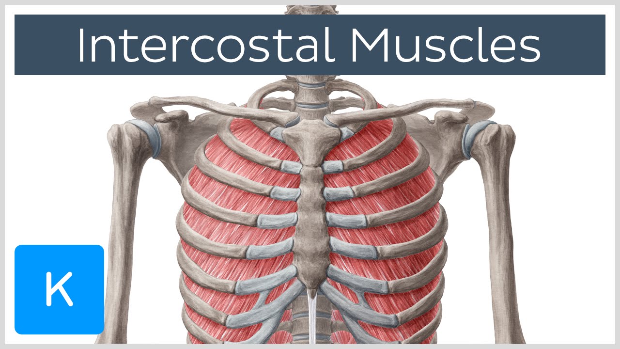
The two muscles which comprise the intermediate muscle group are the serratus posterior inferior, and the serratus posterior superior. Diversitech condensate pump wiring diagram. 3:22 the rib cage is an origin and insertion area for many muscles. The ribs are a set of twelve paired bones which form the protective 'cage' of the thorax. Learn vocabulary, terms and more with flashcards, games and other study tools. The function of the rib cage is to filter the blood it receives, processing the blood. Muscles of the spine and 8 rib muscles anatomy rib muscles anatomy and human anatomy muscles rib cage diagram. Please click on the diagram(s) to view larger version. 2006 kia optima belt diagram. Posted on december 22, 2018december 22, 2018. The intercostal muscles allow ribs to move while breathing. The thoracic cage is part of the axial skeleton (also known as the rib cage), and consists of 24 ribs, the sternum, costal cartilage, and the 12 thoracic vertebrae. Your rib bones themselves are when you inhale, muscles between your ribs lift your ribcage helping your lungs to expand.
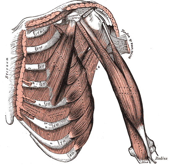
The rib cage is an arrangement of bones in the thorax of all vertebrates except the lamprey. The thoracic cage is part of the axial skeleton (also known as the rib cage), and consists of 24 ribs, the sternum, costal cartilage, and the 12 thoracic vertebrae. Human anatomy for muscle, reproductive, and skeleton. The muscles on your ribcage you are referring to are called the serratus anterior it is a muscle that originates on the surface of the 1st to 8th ribs at the side of the chest and inserts along the entire anterior length of the medial border of th. The primary responsibilities of the ribcage involve protecting the thoracic visceral organs, enclosing the thoracic visceral organs, and is included in the general mechanics of the process of breathing.
Intercostal Muscles Function Area Course Human Anatomy Kenhub Youtube from i.ytimg.com Human anatomy for muscle, reproductive, and skeleton. The function of the rib cage is to filter the blood it receives, processing the blood. As you inhale, the muscles in between the ribs lift the rib cage up, allowing the lungs to expand. 3:27 so let's learn the ribs so we can attach the muscles in the right place. Perform dumbbell pullovers to work the muscles along your rib cage. Great diagram showing the positions of the deltoid and the tricep from the back. The primary responsibilities of the ribcage involve protecting the thoracic visceral organs, enclosing the thoracic visceral organs, and is included in the general mechanics of the process of breathing. This is an online quiz called rib cage muscle diagram.
It is formed by the vertebral column, ribs, and sternum and encloses the heart and lungs.
The thoracic cage makes up the skeleton for the thoracic wall, and provides the attachments needed for the muscles of the neck, thorax. The ribs are a set of twelve paired bones which form the protective 'cage' of the thorax. The two muscles which comprise the intermediate muscle group are the serratus posterior inferior, and the serratus posterior superior. Rib cage diagram with organs. You'll need a bench and one dumbbell to do this exercise. Human anatomy for muscle, reproductive, and skeleton. If you were to develop well defined rib cage muscles, they would give off the appearance of fingers on your sides. Diversitech condensate pump wiring diagram. This post is about rib cage. Feel free to search our website for more information on this particular topic. See more ideas about anatomy, anatomy study, rib cage anatomy. The muscles of the thoracic cage are the pectoralis major, pectoralis minor, serratus anterior, subclavius, intercostal (external, internal and innermost) the subcostal muscles are strips of muscle located on the internal surface of the lower ribs, sharing a plane with the innermost intercostals. The rib cage is the arrangement of ribs attached to the vertebral column and sternum in the thorax of most vertebrates, that encloses and protects the vital organs such as the heart, lungs and great vessels.
Your ribs form a protective cage that encloses many of your delicate internal organs, such as your heart and lungs rib cage muscles. The accompanying diagram reveals the actions of the muscles in this pose.


Posting Komentar
0 Komentar